Theranostics & Dosimetry
Development and evaluation of imaging-guided radionuclide therapies, including quantitative imaging, dosimetry calculations, and characterization of matched diagnostic–therapeutic isotope pairs.
We use quantitative preclinical PET, SPECT and CT to study how radiotracers, antibodies, nanoparticles and lipid-based drug carriers behave inside living systems — supporting theranostics, drug delivery, nanomedicine and translational pharmacology research at UBC and beyond.
The areas below represent some of our most common research applications, but they are far from exhaustive. MIRF specializes in quantitative molecular imaging, combining multi-isotope SPECT, PET and CT with biodistribution, pharmacokinetic, and therapy-efficacy studies to generate robust in vivo data. Because every scan produces quantitative measurements rather than simply anatomical images, we can investigate a wide range of questions related to drug delivery, target engagement, therapeutic response, and biological function in living systems.
Development and evaluation of imaging-guided radionuclide therapies, including quantitative imaging, dosimetry calculations, and characterization of matched diagnostic–therapeutic isotope pairs.
Assessment of therapeutic response through longitudinal imaging, survival studies, and quantitative whole-body biodistribution, enabling treatment effects to be monitored over time within the same animal model.
In vivo tracking of gold, lipid, polymeric, and other nanoparticle-based platforms to characterize biodistribution, clearance pathways, target accumulation, and therapeutic delivery.
Quantitative evaluation of small molecules, biologics, antibodies, peptides, and inhaled therapeutics, supporting preclinical drug-development and translational research programs.
Development of advanced imaging methodologies, including dual-isotope and multi-tracer protocols, quantitative reconstruction, calibration strategies, phantom validation, and image-analysis workflows.
Through a longstanding collaboration with TRIUMF, MIRF plays a leading role in evaluating the in vivo imaging potential of newly available radioisotopes. Many of these non-conventional radionuclides exhibit complex decay characteristics—including multiple gamma emissions and in-growing daughter products—that challenge accurate detection and quantification. MIRF develops and validates the required imaging protocols, including energy-window optimization, calibration, reconstruction, quantitative analysis, and dosimetry, to establish their feasibility for preclinical research. Recent achievements include the first preclinical imaging of 165Er and the first in vivo quantitative imaging studies of 226Ac and 197g/mHg. See the publications →
These examples represent only a subset of the studies supported by MIRF. Our quantitative imaging platform can be adapted to address diverse biological and therapeutic questions across oncology, immunology, neuroscience, cardiology, infectious disease, and drug development. If a molecule, cell, nanoparticle, or therapeutic agent can be labelled, there is a strong possibility it can be tracked, quantified, and studied in vivo.
Static images reveal where a tracer accumulates; dynamic imaging reveals how it moves through the body over time. This SPECT/CT time-course follows the biodistribution of a radiotracer after oral administration, providing a quantitative 4D view of tracer uptake, distribution, and clearance—one of the key advantages of preclinical molecular imaging.
Watch a radiotracer move through the digestive system over time, revealing its transit, distribution, and eventual elimination. Use the controls to replay or navigate through the sequence.
Six flagship studies highlighting the range of MIRF-supported research, including nanomedicine, pulmonary delivery, dual-tracer imaging, targeted alpha therapy, and image-based dosimetry. In each case, quantitative PET/SPECT/CT imaging was central to providing the critical measurement for deriving the biological and therapeutic insights. Each card links to the corresponding publication.
 2025 · Nanomedicine
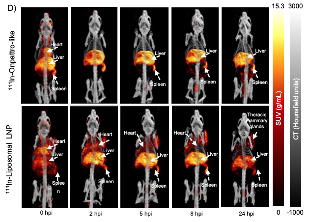
 A new liposomal lipid-nanoparticle design delivers mRNA to tissues beyond the liver. We radiolabelled the particles with 111In and followed them by quantitative SPECT/CT, revealing a roughly 15-fold longer circulation and broader organ uptake than conventional LNPs — cross-validated against ex vivo gamma counting.
 2023 · Theranostic imaging
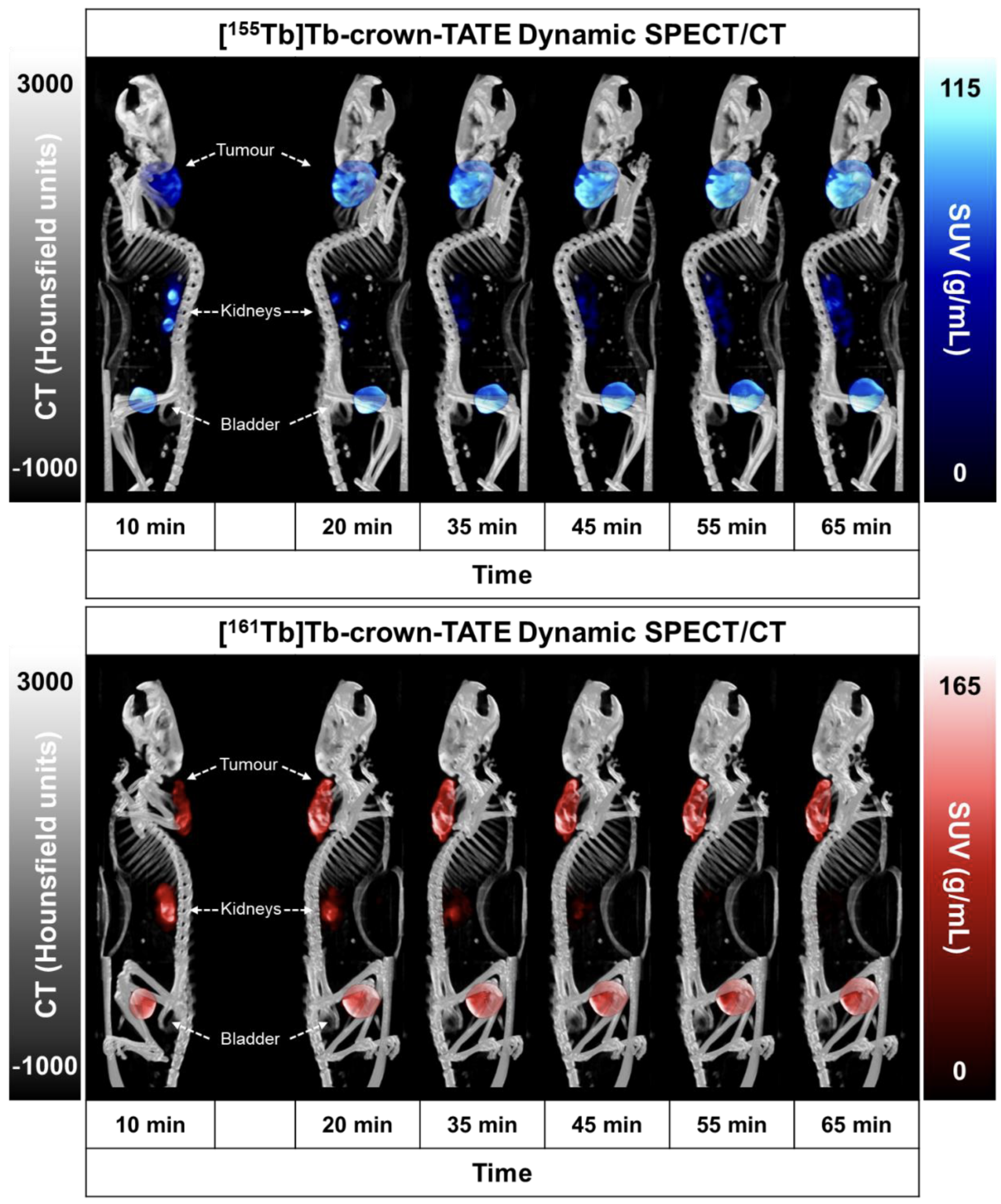
 Crown-TATE labelled with matched terbium isotopes — 155Tb to image, 161Tb to treat. Longitudinal SPECT/CT showed rapid, receptor-specific tumour uptake and that the diagnostic and therapeutic isotopes behave identically, so the scan reliably predicts where the therapy will act.
 2024 · Targeted α-therapy
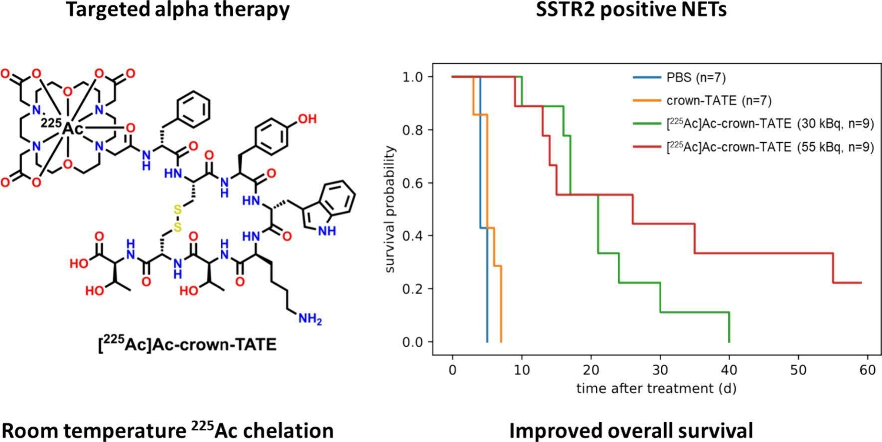
 The same tumour-targeting molecule armed with the alpha-emitter 225Ac. Ex vivo biodistribution and MIRD dosimetry showed the highest absorbed dose went to the tumour, with significantly longer survival in treated mice. The imaging arm comes from its diagnostic partner (the 155Tb pair) — an imaging-guided path to therapy.
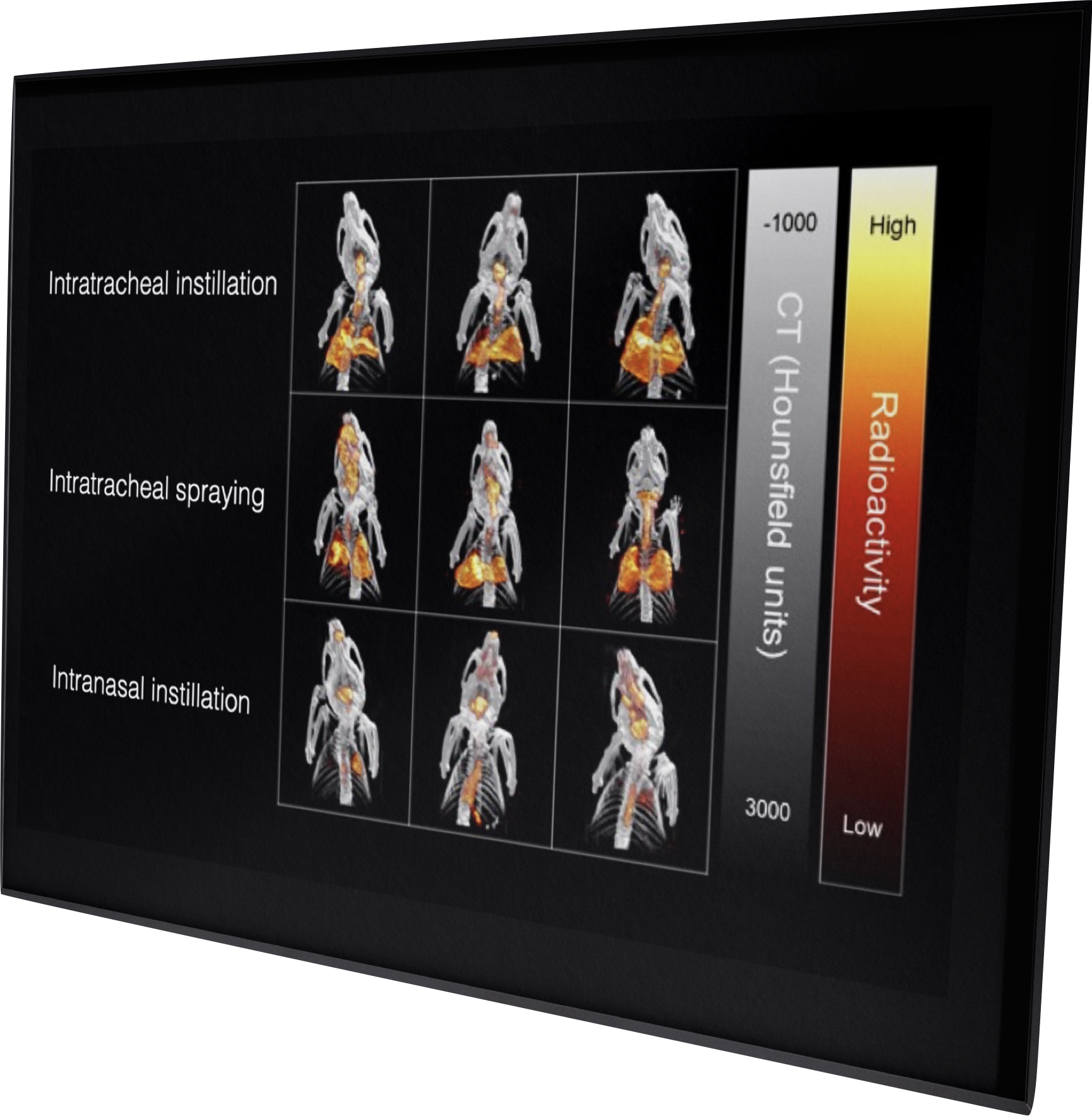
Comparing how inhaled drugs reach the lung is notoriously hard to quantify by conventional analytical methods. By 111In-labelling the nanoparticles and mapping them with quantitative SPECT/CT, we ranked three routes in vivo: intratracheal instillation placed ~95% of the dose in the lungs and spraying ~88%, while intranasal delivery reached only ~29% — the rest ending up in the stomach. The resulting image was a co-winner of the 2020 MILabs Image of the Year Award.
We developed and validated simultaneous dual-isotope (111In + 67Ga) SPECT/CT, tracking two radiopharmaceuticals in the same animal at once — perfectly co-registered in space and time, with quantification errors under 20%. In a proof-of-concept, two co-injected agents’ pharmacokinetics were measured independently in a single mouse.
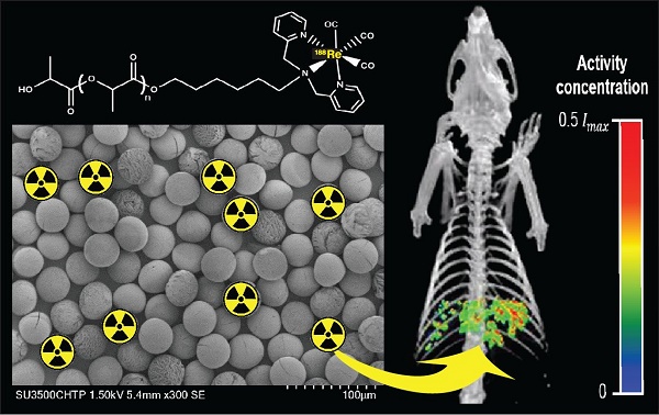 2019 · Dosimetry
 Biodegradable 188Re-labelled microspheres for liver-tumour radioembolization. Because 188Re also emits a gamma photon, quantitative SPECT/CT mapped where the spheres lodged and how long they stayed — producing absorbed-dose maps showing the tumour received three-to-six times the dose of healthy liver. The therapy reports its own dose.
From 197Hg SPECT to 177Lu nanoparticle theranostics, our publications span 2014–2026 across radiochemistry, drug delivery, theranostics and methods development. The full filterable list is one click away.
Browse all publicationsSeparate from our research program, MIRF also runs hands-on training in preclinical PET/SPECT and imaging data analysis — open to graduate students, postdocs and visiting researchers.
Two workshop tracks — an introductory course covering preclinical imaging, the imaging modalities, and the principles of PET, SPECT and CT; and a hands-on imaging data analysis workshop on visualization, quantification and reporting.
We collaborate with academic labs, biotech, radiopharma and translational research groups across Canada and internationally. Whether you need a single experiment or a multi-year project, we'll talk through the right approach.