Integrated PET, SPECT, and CT
in a single preclinical imaging platform.
The MILabs VECTor/CT is a preclinical imaging system that enables PET, SPECT, and CT acquisition within the same animal and imaging session, supporting multimodal molecular and anatomical imaging across a broad range of radiotracers. The system is designed for quantitative small-animal imaging workflows, including sub-millimeter spatial resolution for high-resolution preclinical studies.
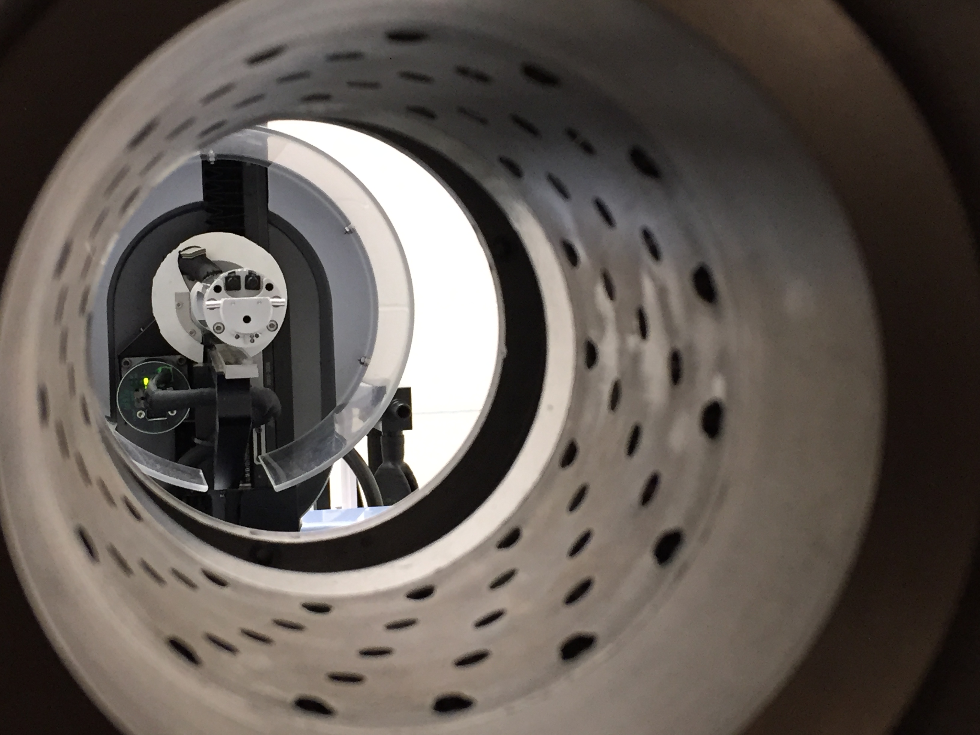
View through the multi-pinhole collimator of the VECTor/CT. The animal bed moves through this collimator during imaging; the triangular detector array (see below) remains stationary.
Molecular and anatomical data, without moving the animal
Running every modality on one bed in a single session means functional and structural data line up automatically — so you can see where a tracer goes and what it does, without the registration headaches and extra animals that separate scanners demand.
Inside the VECTor/CT
Hover over the parts of the scanner to see what each one does. From detector arrays to animal bed.
Ten applications, one scanner
From radiotracer development to gene therapy research, the VECTor/CT supports a broad span of preclinical imaging — connected by a shared infrastructure built for groups whose research moves between modalities, isotopes, and species.
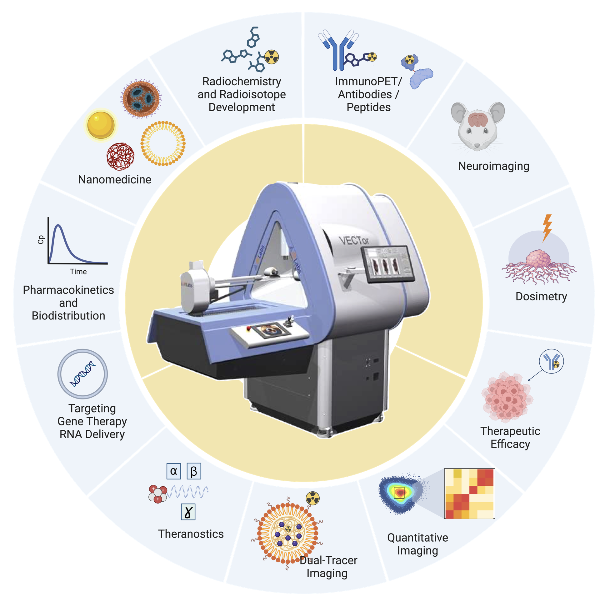
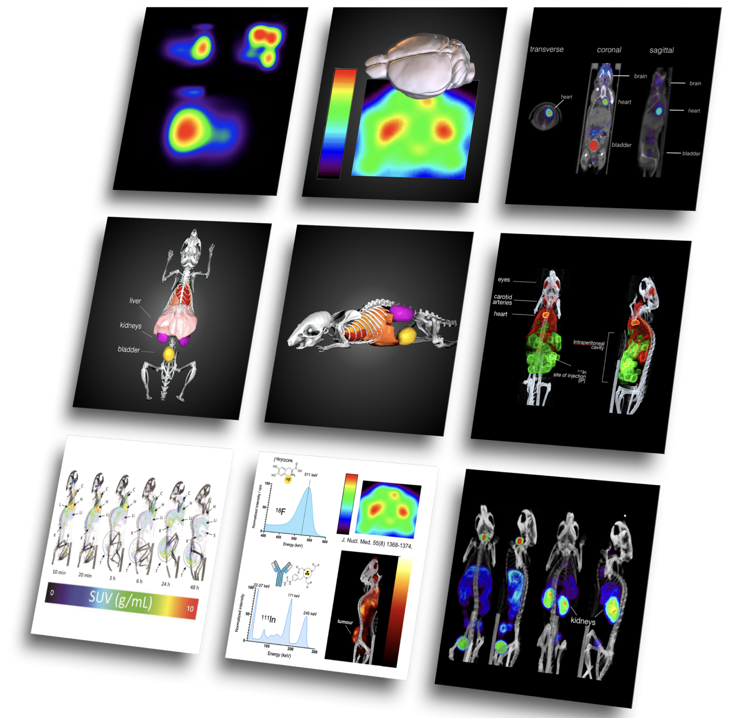
Left: application areas supported by the MIRF VECTor/CT scanner. Right: representative imaging outputs from MIRF studies and collaborations — from quantitative whole-body distributions to detailed tracer-uptake comparisons. For deeper context on specific studies, see our Research page →
Image credits. Composite figure © Cristina Rodríguez, MIRF (UBC). Individual image data acquired at MIRF; specific datasets credited within their originating publications where applicable.
What this means for your work
The same scanner serves two very different audiences. Pick the path that matches your study.
Contract preclinical imaging
Quantitative imaging support for drug discovery, biodistribution, pharmacokinetics, and IND-enabling studies — handled end-to-end by an experienced team.
- Biodistribution and pharmacokinetic profiling
- Tracer radiolabeling via TRIUMF & UBC Pharm Sci partners
- Optional histology & pathology follow-up
- Quantitative reports for R&D or regulatory submissions
- Confidentiality agreements and IP protection in place
Collaborative imaging studies
Full support for academic research — from method development to grant-ready data. Open to UBC, BC-wide, and external collaborators.
- Dual-isotope SPECT and novel tracer development
- Animal ethics protocols managed on our side
- On-site rabbit line for short-lived isotope studies
- Co-authorship and grant collaboration welcomed
- Custom data analysis & visualization tools built in-house
Specs, collimators, and isotopes
Open any section to see the technical detail. For specific questions not covered here, just contact us.
Collimator options
The VECTor/CT supports four interchangeable collimators, each optimized for different combinations of animal size, isotope energy, resolution, and sensitivity. The right choice depends on the study.
| Name | Animal | Best for | Resolution | Sensitivity | Energy |
|---|---|---|---|---|---|
| GPM | Mouse | High-resolution imaging, low-energy isotopes | 0.4 mm | >1500 cps/MBq | Low (< 350 keV) |
| UHR | Rat & mouse | High-resolution rat and mouse imaging | 0.8 mm | >700 cps/MBq | Low (< 350 keV) |
| HE-UHR | Mouse | High-energy isotopes — PET imaging | <0.75 mm 18F · <0.55 mm 99mTc | >6000 cps/MBq 18F · >3000 99mTc | High (> 350 keV) |
| UHS | Mouse | Low-energy isotopes, low injected doses, low count rates | 1.0 mm | >12500 cps/MBq | Low (< 350 keV) |
The table above lists each collimator’s routine resolution; the figures here are the system’s best under ultra-high-resolution, high-count conditions. Combined PET/SPECT workflows run at <0.75 mm. Sources: MILabs · Goorden et al., J Nucl Med 2013;54(2):306.
Source: MILabs CCM-VECTor collimator specifications, calibrated and verified at MIRF.
Compatible radioisotopes
The VECTor/CT supports a wide range of preclinical isotopes — both routine and specialized. Below is a representative list. For isotopes not shown, contact us directly to confirm compatibility.
Multimodal acquisition
PET, SPECT, and CT are all acquired on the same animal in the same session. The CT provides anatomical reference and attenuation correction; PET and SPECT provide functional and molecular information.
Acquisitions are spatially co-registered automatically — no manual alignment needed. This is particularly valuable for studies requiring precise organ localization or for combining quantitative PET/SPECT with anatomical CT context.
The VECTor is among the few preclinical scanners capable of simultaneous PET and SPECT — a unique capability for studies that need to track two separate tracers, or compare PET and SPECT tracers in the same animal.
Animal models supported
Mouse and rat imaging are both supported, with collimator configurations optimized for each.
Animal ethics protocols are managed on the MIRF side for most collaborative studies — reducing the administrative load on the partner team.
Data, analysis, and deliverables
Raw data are reconstructed and quantified using the MILabs software suite, with custom analysis and visualization tools developed in-house at MIRF for specialized applications.
Standard deliverables include reconstructed image volumes, quantitative time-activity curves, regional ROI analysis, and summary figures suitable for publication or regulatory submission.
Optional: histology & pathology assessment.
Tell us what you are trying to image — a drug, a tracer, a disease model — and we will help you figure out the right protocol. 30-minute scoping calls are free.