Nuclear Molecular Imaging.
The Essentials.
A short primer on the techniques used at MIRF — what PET and SPECT actually do, how collimators shape SPECT images, and what makes simultaneous PET/SPECT imaging unique.
What is Nuclear Molecular Imaging?
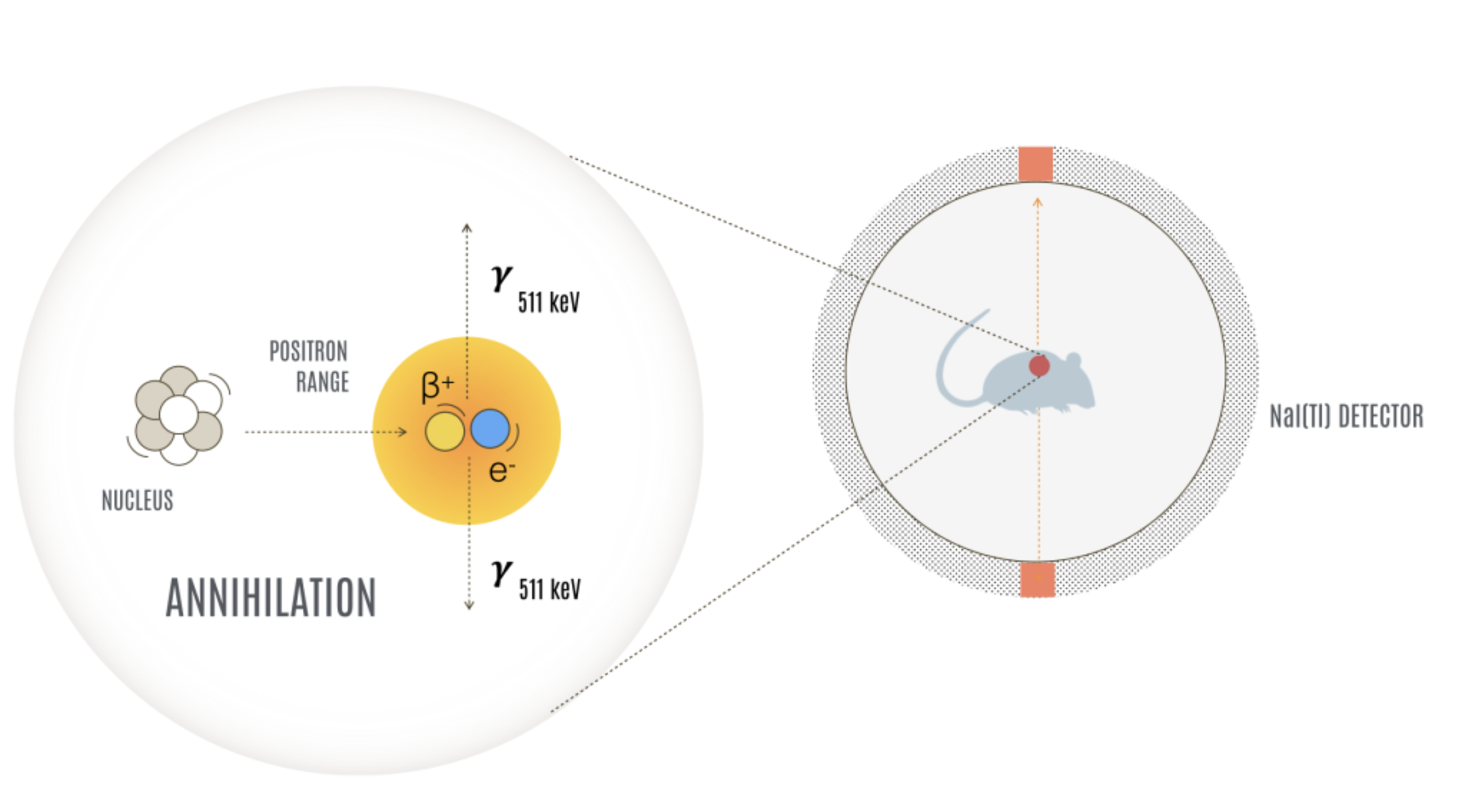
Nuclear molecular imaging uses small amounts of radioactive tracers to visualize biological processes in living animals. Unlike CT or MRI, which primarily depict anatomy, PET and SPECT provide functional information by showing where a tracer accumulates, how tissues metabolize substrates such as glucose, or whether specific receptors are being targeted.
PET and SPECT are highly sensitive techniques that can provide quantitative or semi-quantitative measurements of tracer distribution in vivo. Because imaging is non-invasive, the same animal can be studied repeatedly over time, enabling longitudinal assessment throughout disease progression, treatment response, or tracer evaluation. These capabilities make PET and SPECT important tools in translational imaging, drug development, and radiotracer research.
PET and SPECT, side by side
Where PET & SPECT fit in
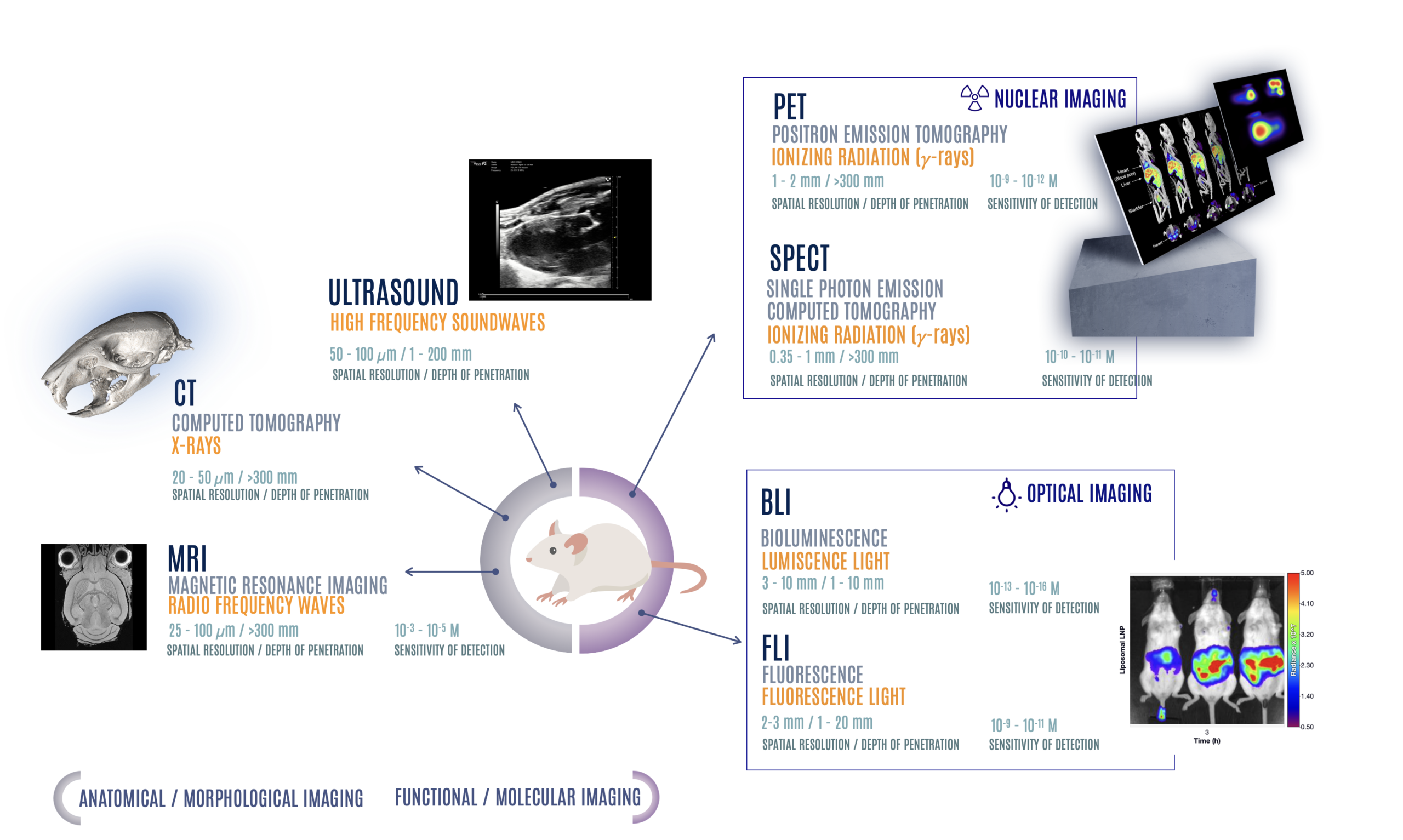
PET and SPECT are part of a broader landscape of preclinical imaging modalities, each with distinct strengths in sensitivity, spatial resolution, tissue penetration, and biological specificity. No single modality captures every aspect of biology, and modality selection depends on the scientific question being asked.
Structural techniques such as CT and MRI provide detailed anatomical information, while molecular imaging approaches reveal functional, metabolic, or cellular processes in vivo. Combining modalities often provides complementary information that cannot be obtained from a single technique alone.
How collimators shape the image
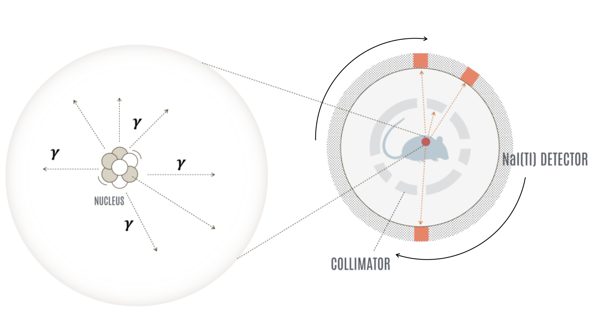
PET does not require physical collimators because coincidence detection electronically constrains the origin of detected photons. SPECT is different: gamma photons are emitted individually and isotropically, meaning their direction cannot be inferred directly from detection alone. In preclinical SPECT, spatial resolution is commonly recovered using multi-pinhole collimators.
A collimator is a dense metal structure — typically made of tungsten — containing precisely machined pinholes. Only photons traveling along selected trajectories reach the detector, while the remainder are absorbed. By acquiring projections from multiple angles and reconstructing them computationally, the scanner generates a three-dimensional map of tracer distribution.
Pinhole geometry introduces an important trade-off between spatial resolution and sensitivity. Smaller pinholes improve image sharpness but allow fewer photons to reach the detector, reducing count sensitivity. Larger pinholes increase photon collection at the expense of spatial resolution.
Modern preclinical SPECT systems address this limitation using multi-pinhole collimators, in which multiple small apertures are arranged in optimized geometries. This approach improves sensitivity while preserving the sub-millimeter spatial resolution achievable in preclinical imaging. Pinhole geometry also introduces magnification, further contributing to high-resolution imaging performance.
More photons, blurrier images.
When to use PET vs SPECT
Why simultaneous PET and SPECT matters
Most preclinical imaging systems are designed for either PET or SPECT acquisition. Simultaneous PET/SPECT imaging within the same animal and imaging session is less common, but provides important experimental advantages by enabling complementary molecular information to be acquired under matched biological conditions.
Two tracers in one animal. PET and SPECT tracers can be evaluated simultaneously within the same biological system, reducing inter-animal variability and enabling direct comparison between complementary molecular pathways, reference tracers, or candidate radiopharmaceuticals.
Co-registered multimodal imaging. Anatomical CT, functional PET, and molecular SPECT data can be acquired within a single imaging workflow, minimizing spatial misregistration and reducing variability associated with repositioning or sequential acquisitions.
Reduced animal usage and improved longitudinal consistency. Multiple imaging readouts can be obtained from the same subject and imaging session, improving statistical consistency while reducing cohort size and experimental burden.